
If you are looking for a refreshing way to improve your health and wellbeing. NDIS hydrotherapy services may be just what you need. Hydrotherapy is also known as aquatic therapy. Offers a unique approach to rehabilitation and pain management that can benefit people of all ages and abilities.
In this blog, we’ll explore the benefits of hydrotherapy services and how they can help you achieve your health goals.
NDIS hydrotherapy services provide individuals access to therapeutic exercise and activities in a warm water pool. Under the guidance of a qualified physiotherapist, participants can engage in a variety of exercises aimed at improving strength, mobility, and flexibility.
Why Choose Hydrotherapy: Here are a few reasons:
- Buoyancy: Water reduces the impact on joints, making exercise easier and less painful, especially for those with conditions like arthritis or joint pain.
- Resistance: The natural resistance of water helps strengthen muscles without the need for heavy equipment.
- Relaxation: The warmth and support of water promote relaxation, reducing stress and tension.
- Improve Circulation: Hydrostatic pressure improves circulation, reducing swelling and adding to tissue healing.
In addition to hydrotherapy, NDIS hydrotherapy services in Altona offer a range of rehabilitation and well-being services tailored to an individual’s needs. Clinical physiotherapists in Altona work closely with participants to develop personalized treatment plans to address specific goals and actions!
NDIS Support for Improved Health and Wellbeing
The NDIS offers a variety of supports and services aimed at improving the overall health and well-being of participants. These supports may include:
- Hydrotherapy: Hydrotherapy, or aquatic therapy, offers a gentle yet effective way for NDIS participants to improve flexibility, or reduce pain and enhance relaxation, through water-based exercise.
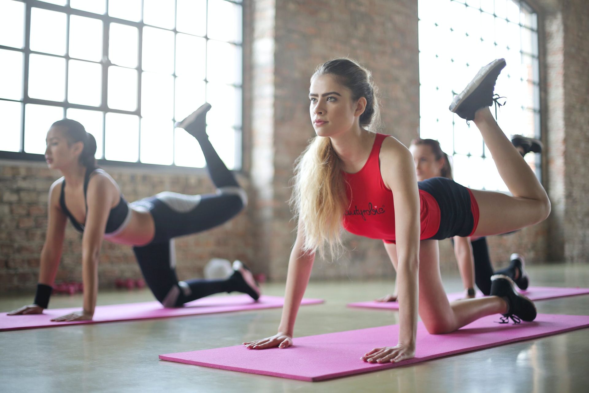
- Exercise Program: NDIS participants can access tailored exercise programs to improve physical fitness, strength, and mobility, promoting overall health and well-being.
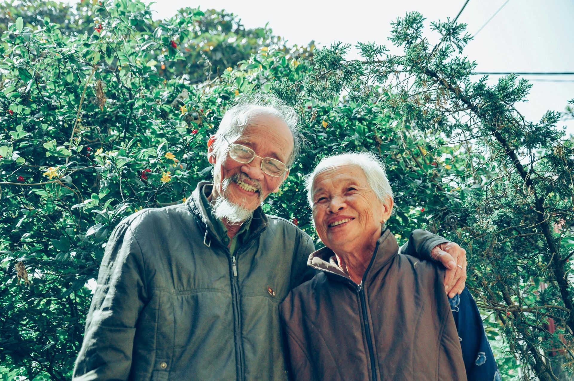
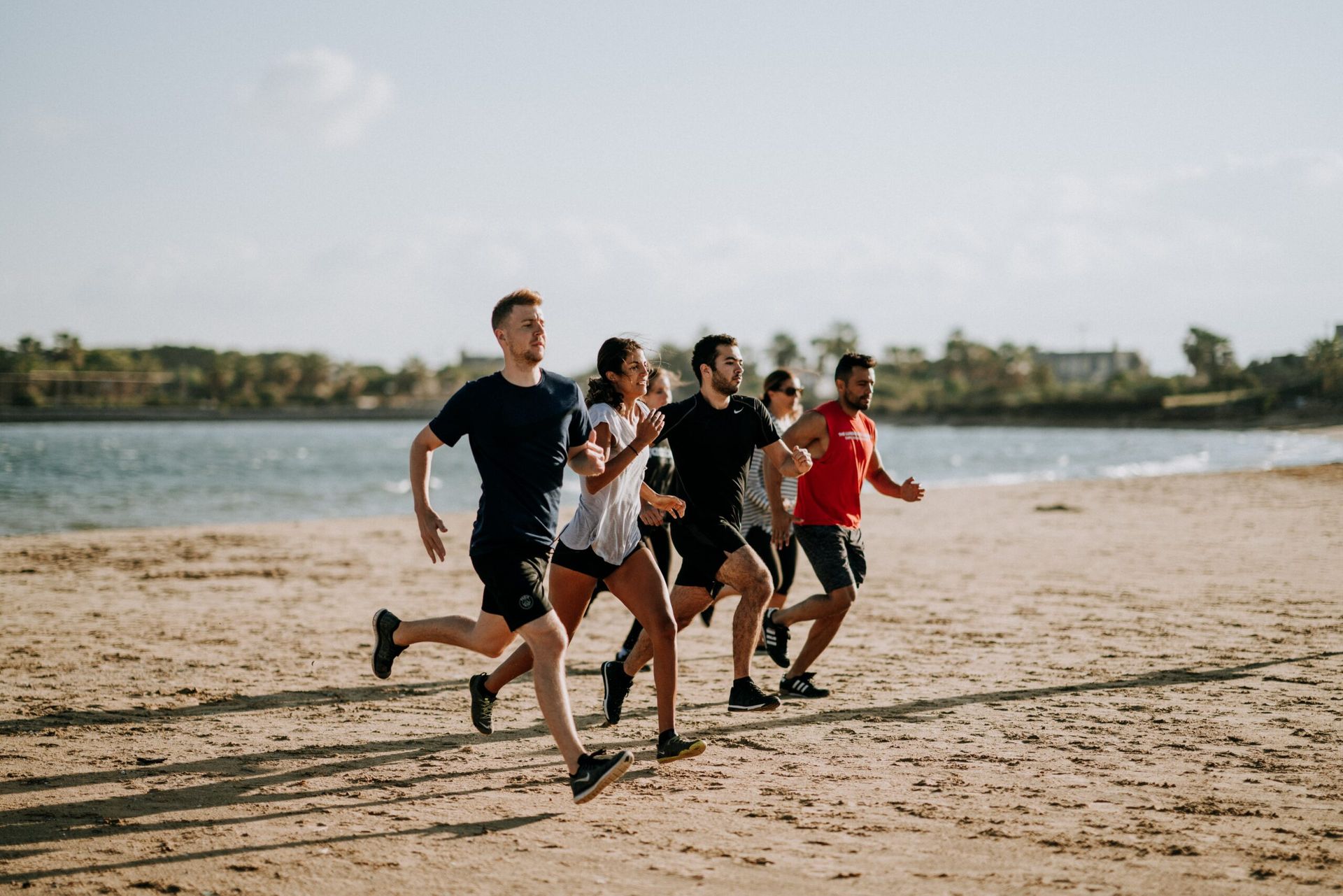
- Outdoor Activity: Engaging in outdoor activities such as hiking, cycling, and nature walks can promote mental well-being, social interaction, and overall happiness for NDIS participants.
- Clinical Physiotherapy: Clinical physiotherapy services are available to NDIS participants to address musculoskeletal issues, improve mobility, and enhance physical function, promoting independence and overall well-being.
Suppose you are looking for a fun and effective way to improve your health and well-being. Consider exploring NDIS hydrotherapy services. Whether you are seeking relief from pain, improving mobility, or simply looking to stay active, hydrotherapy offers a gentle and enjoyable approach to achieving your goals. Combined with NDIS physiotherapy services in Altona, you can access comprehensive care and support to help you on your journey to better health!